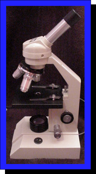
38 light microscope with labels
Compound Light Microscope: Everything You Need to Know A fluorescence microscope, also called a confocal microscope, is a kind of biological microscope that operates by using different light colors and wavelengths over-dyed specimen samples in order for the dye to interact with the light, after which the resulting image is scanned. Fluorescence Microscopy - Explanation and Labelled Images A fluorescence microscope is used to study organic and inorganic samples. Fluorescence microscopy uses fluorescence and phosphorescence to examine the structural organization, spatial distribution of samples. It is particularly used to study samples that are complex and cannot be examined under conventional transmitted-light microscope.
Label a microscope - Teaching resources - Wordwall Label a microscope - Label the Light Microscope - Label the Light Microscope - Microscope slide - label the parts - Year 7 A Microscope.
Light microscope with labels
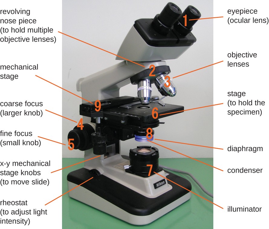
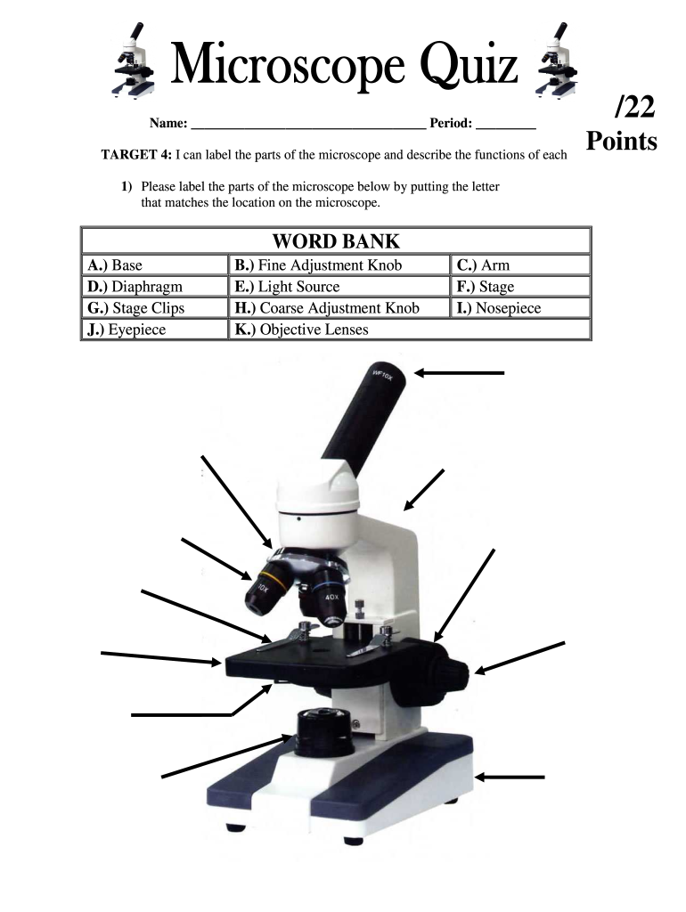
Labs distributes PCR... Welcome to Light Labs. Since 2002, Light Labs has distributed high quality laboratory consumables and equipment, including MultiMax Barrier tips, PCR tubes and strip tubes, PCR plates, and much more. With an emphasis on customer service, we have successfully served the research marketplace with a wide array of laboratory goods. › products › microscopeLAS X Industry Microscope software for Industry | Products ... The software can handle multiple users who have different levels of microscope skills and diverse tasks to accomplish. Profiles according to user’s skills. The LAS X software enables you to create profiles according to the skills and tasks of individual users – from microscopy beginner to expert. It helps you to get reliable results. PDF Parts of the Light Microscope - Science Spot Supports the MICROSCOPE D. STAGE CLIPS HOLD the slide in place C. OBJECTIVE LENSES Magnification ranges from 10 X to 40 X F. LIGHT SOURCE Projects light UPWARDS through the diaphragm, the SPECIMEN, and the LENSES H. DIAPHRAGM Regulates the amount of LIGHT on the specimen E. STAGE Supports the SLIDE being viewed K. ARM Used to SUPPORT the
Light microscope with labels. Compound Light Microscope Labelling Quiz - PurposeGames.com This is an online quiz called Compound Light Microscope Labelling There is a printable worksheet available for download here so you can take the quiz with pen and paper. Your Skills & Rank Total Points 0 Get started! Today's Rank -- 0 Today 's Points One of us! Game Points 15 You need to get 100% to score the 15 points available Actions › products › microscopeMicroscope Objective Lens | Products | Leica Microsystems The objective lens is a critical part of the microscope optics. The microscope objective is positioned near the sample, specimen, or object being observed. It has a very important role in imaging, as it forms the first magnified image of the sample. The numerical aperture (NA) of the objective indicates its ability to gather light and largely determines the microscope’s resolution, the ... Compound Microscope Labeled Diagram | Quizlet High-power objective lense (Magnification- the bigger one) contains the lenses with higher power of magnification Stage clips holds slides on stage Light source Projects light upwards through the diaphragm to allow you to see the specimen Diaphram Disk under the stage that allows light through. Base Bottom or lower part of the microscope Objectives Labeling the Parts of the Microscope Labeling the Parts of the Microscope This activity has been designed for use in homes and schools. Each microscope layout (both blank and the version with answers) are available as PDF downloads. You can view a more in-depth review of each part of the microscope here. Download the Label the Parts of the Microscope PDF printable version here.
Student's Guide: How to Use a Light Microscope An ultraviolet microscope uses UV light to view specimens at a resolution that isn't possible with the common brightfield microscope. It utilizes UV optics, light sources, as well as cameras. Because of the shorter wavelengths of UV light (180-400 nm), the image produced is clearer and more distinct at a magnification approximately double what ... Microscope With Labels Clip Art at Clker.com PEOPLE GOT HERE BY SEARCHING: diagrams of the microscope · light microscope and label · the compound microscope drawing · diagram of microscope with labelling ... proscitech.com.auProSciTech Laboratory supplies and Lab equipment for Histology, Pathology, Light Microscopy, Electron Microscopy and specialist researchers. rsscience.com › stereo-microscopeParts of Stereo Microscope (Dissecting microscope) – labeled ... Labeled part diagram of a stereo microscope Major structural parts of a stereo microscope. There are three major structural parts of a stereo microscope. The viewing Head includes the upper part of the microscope, which houses the most critical optical components, including the eyepiece, objective lens, and light source of the microscope.
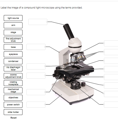
Compound Microscope - Diagram (Parts labelled), Principle and Uses Also called as binocular microscope or compound light microscope, it is a remarkable magnification tool that employs a combination of lenses to magnify the image of a sample that is not visible to the naked eye. Compound microscopes find most use in cases where the magnification required is of the higher order (40 - 1000x). Compound Microscope Parts – Labeled Diagram and their Functions The eyepiece (or ocular lens) is the lens part at the top of a microscope that the viewer looks through. The standard eyepiece has a magnification of 10x. You may exchange with an optional eyepiece ranging from 5x - 30x. [In this figure] The structure inside an eyepiece. The current design of the eyepiece is no longer a single convex lens. Solved Label the image of a compound light microscope using - Chegg Expert Answer. 100% (17 ratings) Transcribed image text: Label the image of a compound light microscope using the terms provided. Microscope Types (with labeled diagrams) and Functions This is an advanced microscope that has specific application in viewing, observing and measuring the optical thickness and phase of completely transparent specimens and objects. A tiny interferometer is used and a specimen is placed on beam path of it. This path is split and then rejoined to create two superimposed images of the specimen in focus.
Instruments of Microscopy | Microbiology | | Course Hero
Parts of the Microscope with Labeling (also Free Printouts) Parts of the Microscope with Labeling (also Free Printouts) By Editorial Team March 7, 2022 A microscope is one of the invaluable tools in the laboratory setting. It is used to observe things that cannot be seen by the naked eye. Table of Contents 1. Eyepiece 2. Body tube/Head 3. Turret/Nose piece 4. Objective lenses 5. Knobs (fine and coarse) 6.

Simple Microscope - Diagram (Parts labelled), Principle ...
Microscope With Labels clip art - Pinterest Jul 3, 2012 - Download Clker's Microscope With Labels clip art and related ... Optical Microscope, Microscopes, Focus Light, Industrial Machine, Things.

Free Microscope Drawing, Download Free Microscope Drawing png ...
Light microscopes - Cell structure - Edexcel - BBC Bitesize Microscopes are used to produce magnified images. There are two main types of microscope: light microscopes are used to study living cells and for regular use when relatively low magnification and...

Compound and Stereo- microscopes - Microscopes 4 Schools
Microscope Labeling Game - PurposeGames.com This is an online quiz called Microscope Labeling Game There is a printable worksheet available for download here so you can take the quiz with pen and paper. This quiz has tags. Click on the tags below to find other quizzes on the same subject. Science microsope Your Skills & Rank Total Points 0 Get started! Today's Rank -- 0 's Points 15 Actions

Microscope Diagram Labeled, Unlabeled and Blank | Parts of a ...
Label the microscope - Science Learning Hub All microscopes share features in common. In this interactive, you can label the different parts of a microscope. Use this with the Microscope parts activity to help students identify and label the main parts of a microscope and then describe their functions. Drag and drop the text labels onto the microscope diagram.
Solved PLEASE HELP THANK YOU- LIGHT SOURCE WILL BE NUMBER 1 ...
› microscopy › enZEISS Elyra 7 with Lattice SIM² Super-Resolution Microscope Lattice SIM² comes with outstanding out-of-focus light suppression, giving you the sharpest sectioning in widefield microscopy even for highly scattering samples. SIM² image reconstruction robustly reconstructs all structured-illumination-based acquisition data of your Elyra 7 – with minimal artefacts – for living and fixed samples.
Label the Microscope by Crista Tiboldo | Teachers Pay Teachers
Simple Microscope - Diagram (Parts labelled), Principle, Formula ... Feb 23, 2022 ... Dating back to the 14th century, simple microscope is the most basic of the various microscopes available. It is a type of optical ...

Parts of a microscope with functions and labeled diagram
Addgene: Using a Light Microscope Protocol It is best practice to start with the lowest power objective to find your sample. Place your slide (or other sample type) on the microscope stage. If using a slide, you can secure it into place using the metal clips on the stage. Turn on the power source and use the stage arm to move the stage so that the light shines onto your sample.

Compound Microscope – Diagram (Parts labelled), Principle and ...
Labelled Diagram Of A Light Microscope | Products & Suppliers There are many applications for solar simulators and light simulators. Some products are designed to test coatings and paints, paper and labels, optical ...

Parts of a Microscope - SmartSchool Systems
Light Microscope: Functions, Parts and How to Use It To use a light microscope, you can follow the steps below carefully. Start with a low lens and a clean slide. The microscope stage should be lowered as low as possible. Center the slide so that the specimen is under the objective lens. Use the coarse adjustment knob to get a general focus. Then slowly move up the stage until focus is achieved.

Compound Microscope Parts – Labeled Diagram and their ...
Parts of a microscope with functions and labeled diagram Microscopic illuminator - This is the microscopes light source, located at the base. It is used instead of a mirror. It captures light from an external source of a low voltage of about 100v. Condenser - These are lenses that are used to collect and focus light from the illuminator into the specimen.

Microscope Label Diagram | Quizlet
A Study of the Microscope and its Functions With a Labeled Diagram ... To better understand the structure and function of a microscope, we need to take a look at the labeled microscope diagrams of the compound and electron microscope. These diagrams clearly explain the functioning of the microscopes along with their respective parts. Man's curiosity has led to great inventions. The microscope is one of them.
simple light microscope labeled - Clip Art Library
Label the Light Microscope - Labelled diagram - Wordwall Drag and drop the pins to their correct place on the image.. Eyepiece, Light Source, Base, Stage, Stage Clips, Fine Focus, Coarse Focus, Arm, Objective Lens.
Transmitted light microscope B3 Professional series B3-220ASC ...
Light Microscope- Definition, Principle, Types, Parts, Labeled Diagram ... A light microscope is a biology laboratory instrument or tool, that uses visible light to detect and magnify very small objects and enlarge them. They use lenses to focus light on the specimen, magnifying it thus producing an image. The specimen is normally placed close to the microscopic lens.

Light Microscopy | BioNinja
› microscopy › enZEISS Lattice Lightsheet 7 The importance of gentle light sheet imaging at high resolution cannot be overestimated for the study of subcellular processes. With Lattice Lightsheet 7, ZEISS makes access to the benefits of this advanced technology amazingly simple.
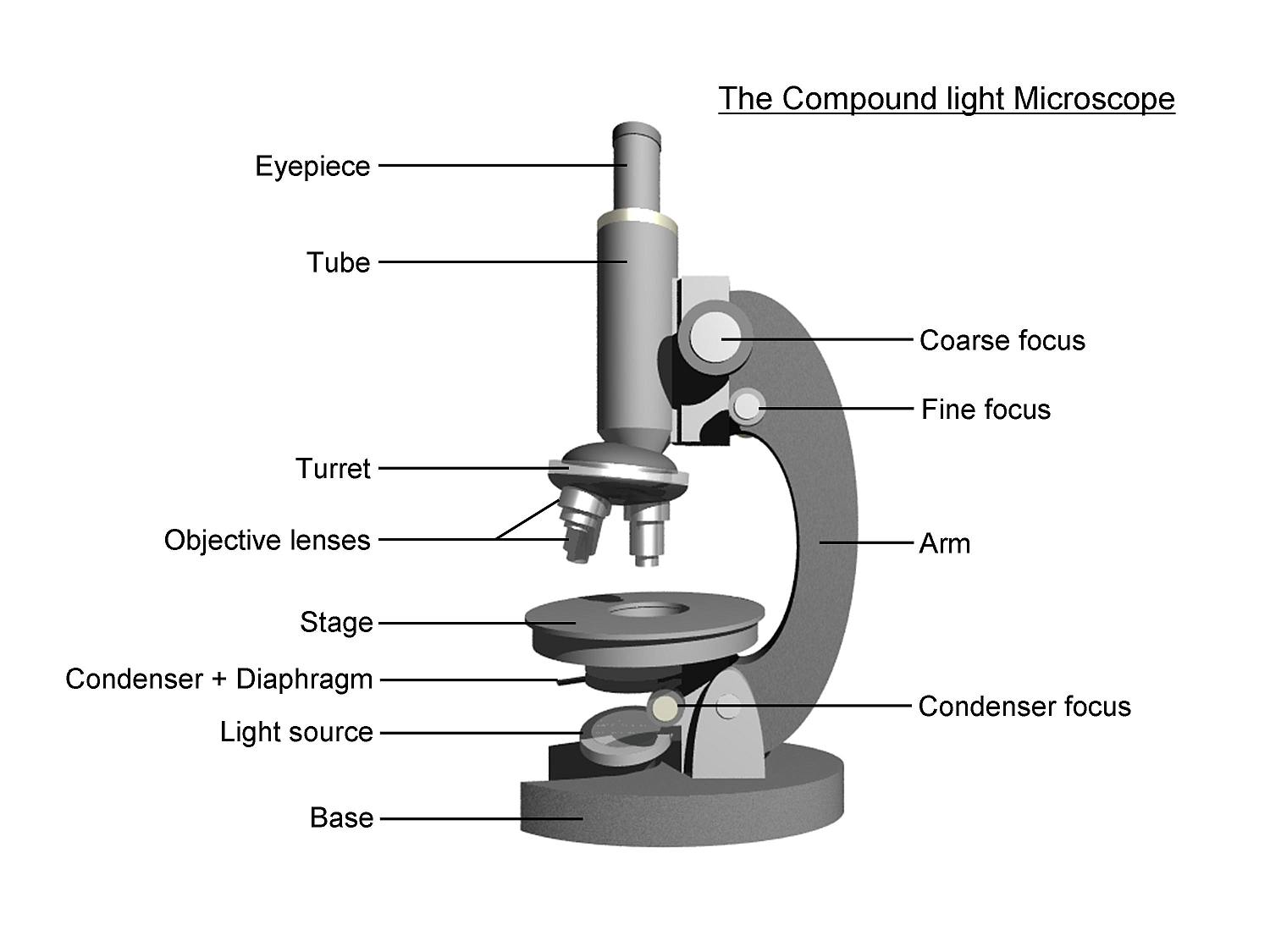
The Compound Light Microscope
Labeling the Parts of the Microscope - Pinterest Jan 13, 2016 - Free worksheets for labeling parts of the microscope including a worksheet that is ... Microscope, Assessment, Labels, Science, Education,.

Photo Compound microscope with labels Image #3850568
Sperm Under Microscope with Labeled Diagram - AnatomyLearner Under the light microscope, the sperm consists of two main portions - the head and the tail. But, the electron microscope shows four different parts in the tail of spermatozoa. ... So, this article provides the details structural features of sperm under the light microscope. All the labeled diagrams might help you identify the sperms from ...

label microscope diagram | Charts | Microscope, Anatomy bones ...
Microscope, Microscope Parts, Labeled Diagram, and Functions Majority of high quality microscopes used in laboratory include an Abbe condenser with an iris diaphragm. When iris diaphragm is combined with Abbe condenser, it control both the quantity of light applied as well as focus on the specimen. Aperture: It is the hole in the stage through which the base (transmitted) light reaches the stage.
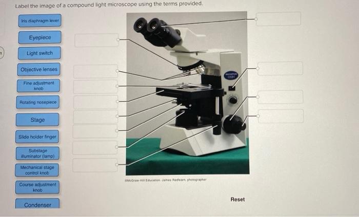
Solved Label the image of a compound light microscope using ...
Light Microscope Parts, Function & Uses - Study.com Anton van Leeuwenhoek (1632-1723) invented a simple (one-lens) microscope around 1670. Leeuwenhoek made lenses by carefully grinding and polishing solid glass to make his microscopes.
Answered: Microscope Structure and Function… | bartleby
Microscope Labeling - The Biology Corner 1) Start with scanning (the shortest objective) and only use the COARSE knob . Once it is focused… 2) Switch to low power (medium) and only use the COARSE knob . You may need to recenter your slide. Once it is focused.. 3) Switch to high power (long objective).

Microscope Diagram and Quiz | Science diagrams, Science ...
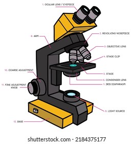
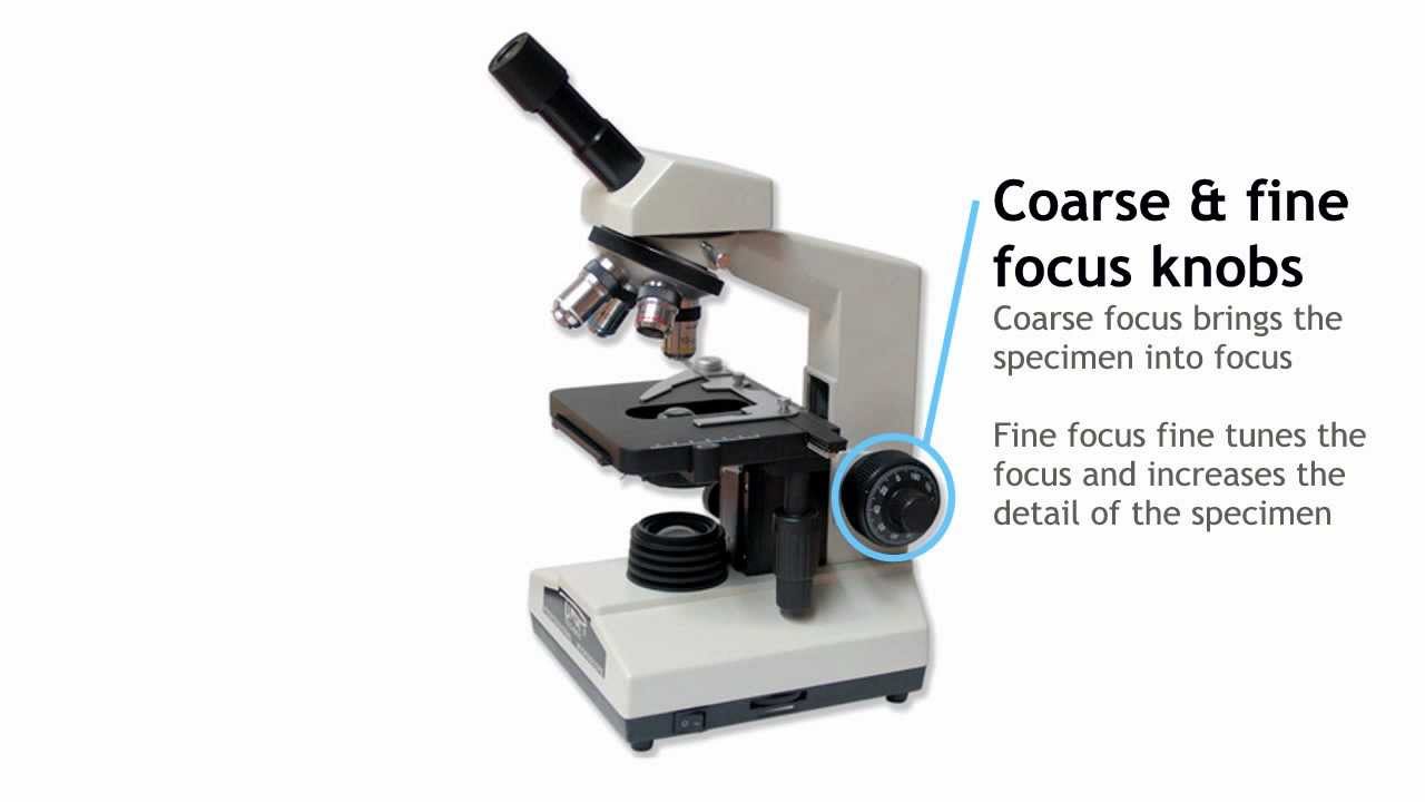
Microscope Parts and Functions Body tube (Head): The body tube connects the eyepiece to the objective lenses. Arm: The arm connects the body tube to the base of the microscope. Coarse adjustment: Brings the specimen into general focus. Fine adjustment: Fine tunes the focus and increases the detail of the specimen. Nosepiece: A rotating turret that houses the objective lenses.

Light Microscope- Definition, Principle, Types, Parts ...
Compound Microscope Parts, Functions, and Labeled Diagram Compound Microscope Definitions for Labels. Eyepiece (ocular lens) with or without Pointer: The part that is looked through at the top of the compound microscope. Eyepieces typically have a magnification between 5x & 30x. Monocular or Binocular Head: Structural support that holds & connects the eyepieces to the objective lenses.
40,724 Light microscope Images, Stock Photos & Vectors ...
PDF Parts of the Light Microscope - Science Spot Supports the MICROSCOPE D. STAGE CLIPS HOLD the slide in place C. OBJECTIVE LENSES Magnification ranges from 10 X to 40 X F. LIGHT SOURCE Projects light UPWARDS through the diaphragm, the SPECIMEN, and the LENSES H. DIAPHRAGM Regulates the amount of LIGHT on the specimen E. STAGE Supports the SLIDE being viewed K. ARM Used to SUPPORT the

Types of Microscopes | Light vs. Electron Microscope - Video ...
› products › microscopeLAS X Industry Microscope software for Industry | Products ... The software can handle multiple users who have different levels of microscope skills and diverse tasks to accomplish. Profiles according to user’s skills. The LAS X software enables you to create profiles according to the skills and tasks of individual users – from microscopy beginner to expert. It helps you to get reliable results.

Label the light microscope | Teaching Resources
Labs distributes PCR... Welcome to Light Labs. Since 2002, Light Labs has distributed high quality laboratory consumables and equipment, including MultiMax Barrier tips, PCR tubes and strip tubes, PCR plates, and much more. With an emphasis on customer service, we have successfully served the research marketplace with a wide array of laboratory goods.

Compound Microscope Parts – Labeled Diagram and their ...

Labeling the Parts of the Microscope | Microscope World Resources

Labelling a Microscope Diagram | Quizlet
Microscope Fill In The Blank - Fill Online, Printable ...

Label the microscope — Science Learning Hub

Microscope Maintenance Tips | Science supplies, Multi step ...
Parts of a Compound Light Microscope
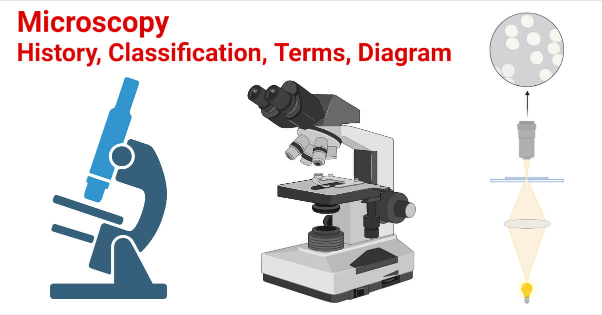
Microscopy- History, Classification, Terms, Diagram

Parts of a microscope with functions and labeled diagram

Compound Light Microscope Labeling Diagram | Quizlet

Monday 10/19/15 AIM: how do the parts of the compound light ...
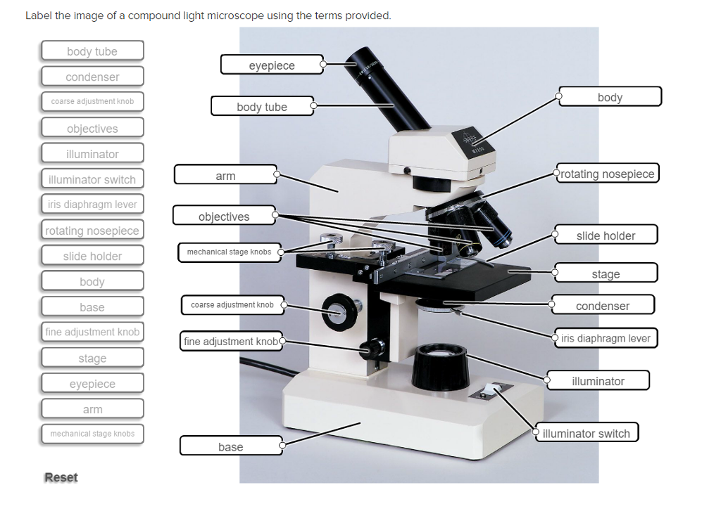
Solved Label the image of a compound light microscope using ...
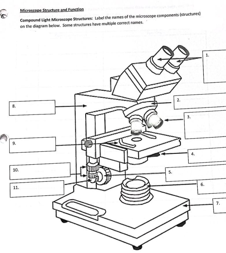
The Compound Light Microscope Label the following parts on ...
Post a Comment for "38 light microscope with labels"