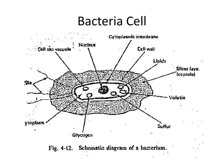
39 bacterial cell picture with labels
Animal Cell Diagram Stock Photos and Images - Alamy Animal cell anatomy on green background. 3D illustration. ID: H9FENY (RF) Plant Cell and Animal cell structure. cross section and anatomy of cell. Biology Chart. Vector illustration on a white background. detailed diagram. ID: 2DHY2W8 (RF) Anatomy of animal cell in three different drawing styles. BYJUS The structure of bacteria is known for its simple body design. Bacteria are single-celled microorganisms with the absence of the nucleus and other c ell organelles; hence, they are classified as prokaryotic organisms. They are also very versatile organisms, surviving in extremely inhospitable conditions. Such organisms are called extremophiles.
600+ Free Bacteria & Virus Images - Pixabay 639 Free images of Bacteria Related Images:virusinfectionhealthcoronavirusdiseasemedicalbiologycovid-19medicine Bacteria and virus high resolution images. Find your perfect picture for your project. 361114 bacteriaillnessvirus 31566 koli bacteria 23073 bacteriamicrobiology 705182 virusmicroscope 30281 coronasprayearth 30570 monsterblueinternet

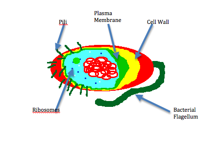
Bacterial cell picture with labels
Bacteria cell diagram Images, Stock Photos & Vectors - Shutterstock Bacteria cell diagram royalty-free images 2,581 bacteria cell diagram stock photos, vectors, and illustrations are available royalty-free. See bacteria cell diagram stock video clips Image type Orientation Artists More Sort by Biology Healthcare and Medical bacterium cell anatomy cell wall virus microbiology eukaryote prokaryotes › articles › s41591/020/01182-9The distribution of cellular turnover in the human body - Nature Jan 11, 2021 · We found that the total turnover rate of the human body is 0.33 ± 0.02 × 10 12 (330 ± 20 billion) cells d −1 (equal to about 4 million cells s −1).About 86% of these cells are blood cells ... Bacteria Diagram Labeled | Cell diagram, Prokaryotic cell, Medical ... 445 views. Find this Pin and more on Biology by Fernanda Newman. Cell Structure. Cell Wall. Korean Words. Science Facts. Microbiology. Study Notes. Medical School.
Bacterial cell picture with labels. Bacterial Cell Structure Labeling Diagram - Quizlet Bacterial Cell Structure Labeling STUDY Learn Flashcards Write Spell Test PLAY Match Gravity Created by adkelly22 Terms in this set (16) Cytoplasm Water-based solution filling the entire cell Ribosomes Tiny particles composed of protein and RNA that are the sites of protein synthesis Nucleoid Composed of condensed DNA molecules. Cell Membrane Animal Cell Labeled Diagram Pictures, Images and Stock Photos Browse 19 animal cell labeled diagram stock photos and images available, or start a new search to explore more stock photos and images. Newest results. ... Labeled educational bacteria internal structure scheme. Biological blue green algae diagram with carboxysome, thylakoid and phycobilisome parts location inside cell. animal cell labeled ... Animal Cell Labeled Pictures, Images and Stock Photos Labeled educational bacteria internal structure scheme. Biological blue green algae diagram with carboxysome, thylakoid and phycobilisome parts location inside cell. Animal cell Cells consist of a protoplasm enclosed within a membrane, which contains many biomolecules such as proteins and nucleic acids. Toxoplasma Gondii Structure Draw a labelled diagram of a bacterial cell. - Careers360 Buy Now NEET Foundation + Knockout NEET 2024 (Easy Installment) Personalized AI Tutor and Adaptive Time Table, Self Study Material, Unlimited Mock Tests and Personalized Analysis Reports, 24x7 Doubt Chat Support,.
Bacteria Labeled Stock Illustrations - Dreamstime Bacteria Labeled Stock Illustrations - 220 Bacteria Labeled Stock Illustrations, Vectors & Clipart - Dreamstime Bacteria Labeled Illustrations & Vectors Most relevant Best selling Latest uploads Within Results People Pricing License Media Properties More Safe Search 220 bacteria labeled illustrations & vectors are available royalty-free. Next page A Labeled Diagram of the Animal Cell and its Organelles A Labeled Diagram of the Animal Cell and its Organelles. There are two types of cells - Prokaryotic and Eucaryotic. Eukaryotic cells are larger, more complex, and have evolved more recently than prokaryotes. Where, prokaryotes are just bacteria and archaea, eukaryotes are literally everything else. From amoebae to earthworms to mushrooms, grass ... Bacterial Staining Microbiology Images ... - Science Prof Online 1. Endospore stain of Bacillus subtilis showing both endospores (green) & vegetative cells (pink) @1000xTM; 2. Negative endospore stain showing only vegetative cells @1000xTM; 3. Malachite green primary staining step of endopore stain with slide being heated over water bath; 4. Applying counterstain (safrinin) to bacterial smear as last step of endospore stain; Endospore stained slide, with ... › content-not-availableError - UpToDate UpToDate, electronic clinical resource tool for physicians and patients that provides information on Adult Primary Care and Internal Medicine, Allergy and Immunology, Cardiovascular Medicine, Emergency Medicine, Endocrinology and Diabetes, Family Medicine, Gastroenterology and Hepatology, Hematology, Infectious Diseases, Nephrology and Hypertension, Neurology, Obstetrics, Gynecology, and Women ...
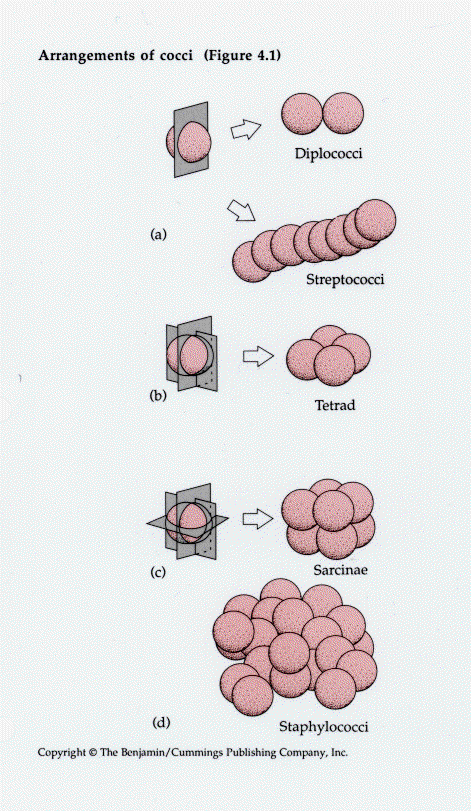
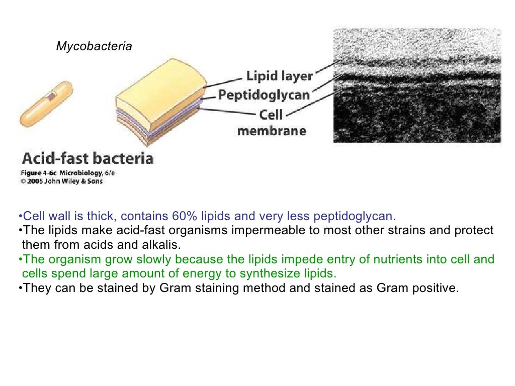
PHOTO GALLERY OF BACTERIA - Microbiology in pictures (the cells stain a weak Gram-negative) Microscopic appearance: Spirochetes: Oxygen relationship: microaerobic: Motility: motile: Catalase test:-Oxidase test:- ... Colonies of various bacteria. Bacteria photos. PICTURE OF THE MONTH. BACTERIA 2013 DECEMBER. DISK DIFFUSION METHOD FOR TESTING OF ANTIBIOTIC SUSCEPTIBILITY OF BACTERIA: Bacteria: Cell Walls - General Microbiology 4 Bacteria: Cell Walls . It is important to note that not all bacteria have a cell wall.Having said that though, it is also important to note that most bacteria (about 90%) have a cell wall and they typically have one of two types: a gram positive cell wall or a gram negative cell wall.. The two different cell wall types can be identified in the lab by a differential stain known as the Gram stain. Different Size, Shape and Arrangement of Bacterial Cells When viewed under light microscope, most bacteria appear in variations of three major shapes: the rod (bacillus), the sphere (coccus) and the spiral type (vibrio). In fact, structure of bacteria has two aspects, arrangement and shape. So far as the arrangement is concerned, it may Paired (diplo), Grape-like clusters (staphylo) or Chains (strepto). › a-to-z-guides › manuka-honeyManuka Honey: Medicinal Uses, Benefits, and Side Effects - WebMD Feb 20, 2021 · Health Research Board: A Picture of Health 2008: "Healing with honey." Jull, A. British Journal of Surgery. 2008; vol 95: pp 175-182. Natural Medicines Comprehensive Database.
Bacteria | CK-12 Foundation
› NeilMed-NeilCleanse-PiercingNeilMed NeilCleanse Piercing Aftercare, Fine Mist, 6.3 Fluid ... Actual product packaging and materials may contain more and/or different information than that shown on our Web site. We recommend that you do not solely rely on the information presented and that you always read labels, warnings, and directions before using or consuming a product.

PHARMA WISDOM: Mechanism of Action of Chemotherapeutic drugs
Bacteria in Microbiology - shapes, structure and diagram Bacterial endospores layers Bacteria cells are the smallest living cells that are known; even though viruses are smaller than bacteria, viruses are not living cells. There are different types of bacteria with various sizes, shapes, and structures. The bacteria shapes, structure, and labeled diagrams are discussed below. Sizes
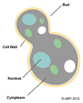
Fungi - ABPI - Resources for Schools
Bacteria Labeled Diagram Stock Vector Image & Art - Alamy Download this stock vector: Bacteria Labeled Diagram - EG0XT7 from Alamy's library of millions of high resolution stock photos, illustrations and vectors.

cell_poster_bacteria_types.JPG 1,182×1,555 pixels | LAB | Pinterest | Poster
Label the Bacterium Cell - EnchantedLearning.com flagellum - A long whip-like structure used for locomotion (movement). Some bacteria have more than one flagellum. pili - (singular is pilus) Hair-like projections that allow bacterial cells to stick to surfaces and transfer DNA to one another. plasma membrane - A permeable membrane located within the cell wall.
American Urological Association - Inverted Papilloma
Bacterial cells - Cell structure - Edexcel - GCSE Combined Science ... Feature Eukaryotic cell (plant and animal cell) Prokaryotic cell (bacterial cell) Size: Most are 5 μm - 100 μm: Most are 0.2 μm - 2.0 μm: Outer layers of cell
PPT - Wastewater Microbiology PowerPoint Presentation - ID:1635113
Structure of Bacterial Cell (With Diagram) - Biology Discussion It is a tough and rigid structure of peptidoglycan with accessory specific materials (e.g. LPS, teichoic acid etc.) surrounding the bacterium like a shell and lies external to the cytoplasmic membrane. It is 10-25 nm in thickness. It gives shape to the cell. Nucleus: The single circular double-stranded chromosome is the bacterial genome.

Fun With Microbiology (What's Buggin' You?): Bacterial Vaginosis
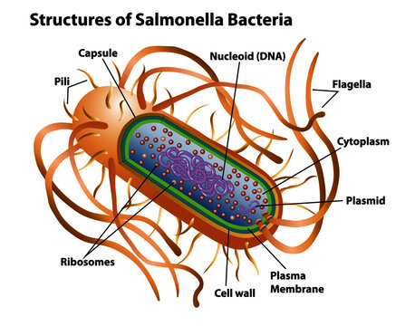
Bacteria Cell Structures with Labels Stock Vector - Dreamstime Get 15 images free trial Bacteria Cell Structures with labels Royalty-Free Vector Bacterial cell structures labeled on a bacillus cell with nucleoid DNA and ribosomes. External structures include the capsule, pili, and flagellum. Morphology of internal structures of bacteria. cell anatomy bacteria, prokaryotic cell, cell, internal structures,

Katy J Negus. BA Hons. CG Arts & Animation: Making an Antibody Research
50 Striking Microscopic Images of Viruses and Bacteria Click through the slideshow above to see 50 striking electron micrographs of some of the world's most dangerous and deadly disease-causing viruses and bacteria. Know your flu risk. Check out the ...
Review
Structure of Bacteria (With Diagram) | Microbiology Bacterial cell wall is extremely thin (10-25 nm thick) and provides rigidity and a definite shape to the cell. 7. Chemically, the cell wall is composed of mucopeptide (murein) scaffolding or platform formed by N- acetyl glucosamine and N-acetyl muramic acid molecules arranged in alternate chains. 8.

Pathology Outlines - Plasma cells
97,783 Bacteria Cell Stock Photos and Images - 123RF Bacteria Cell Stock Photos And Images 97,783 matches Page of 978 Structure of a bacterial cell. Anatomy of the prokaryote. unicellular organism. Vector diagram for your design, educational, medical, biological and science use Bacteria vector icon isolated on transparent background, Bacteria logo concept

Quia - 9AP Chapter 27 - Bacteria and Archaea (detailed)
en.wikipedia.org › wiki › Monoclonal_antibodyMonoclonal antibody - Wikipedia There may also be bacterial contamination and, as a result, endotoxins that are secreted by the bacteria. Depending on the complexity of the media required in cell culture and thus the contaminants, one or the other method (in vivo or in vitro) may be preferable. The sample is first conditioned, or prepared for purification.

Jeff Schneider Teaching Resources | Teachers Pay Teachers
Interactive Bacteria Cell Model - CELLS alive Pili, Fimbriae: These hollow, hairlike structures made of protein allow bacteria to attach to other cells. A specialized pilus, the sex pilus, allows the transfer of plasmid DNA from one bacterial cell to another. Pili (sing., pilus) are also called fimbriae (sing., fimbria). Flagella: The purpose of flagella (sing., flagellum) is motility.
32 Label A Bacterial Cell - Labels Database 2020
Image Library | CDC Online Newsroom | CDC Under a high magnification of 21674X, this digitally-colorized, scanning electron microscopic (SEM) image depicts a view of a dividing, Escherichia coli bacterium, clearly displaying the point at which the bacteria's cell wall was splitting into two separate organisms. See PHIL 7137 for a black and white version of this image.
35 Label Bacteria Cell - Labels 2021
3 Common Bacteria Shapes - ThoughtCo Bacteria Shapes The three basic shapes of bacteria include cocci (blue), bacilli (green), and spirochetes (red). PASIEKA/Science Photo Library/Getty Images By Regina Bailey Updated on August 20, 2019 Bacteria are single-celled, prokaryotic organisms that come in different shapes.
Bacterial Cell
medicalxpress.com › news › 2022-05-success-chimericResearchers seek to improve success of chimeric antigen ... May 12, 2022 · "CAR-T cell therapy is a promising treatment for non-Hodgkin lymphoma, especially for patients who have relapsed or those who have not responded to prior therapies," says Tae Hyun Hwang, Ph.D., a ...
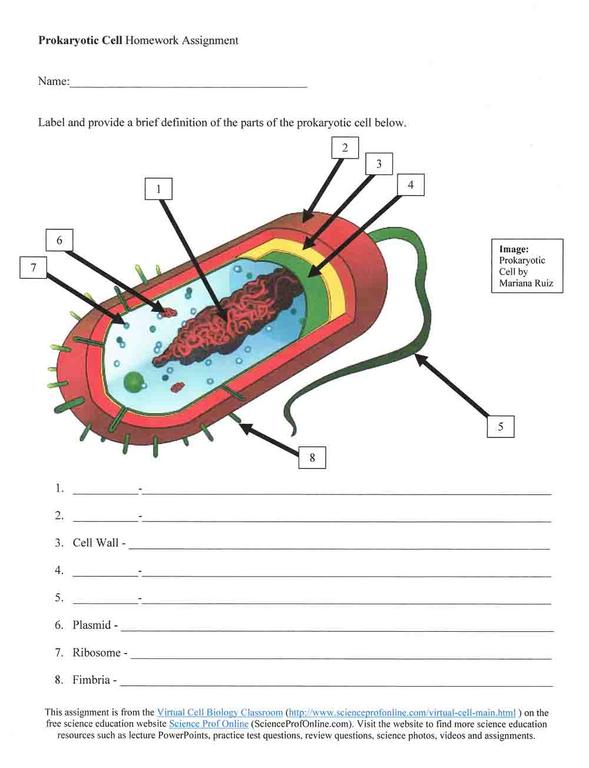
Prokaryotic Cell Diagram Worksheets
medicalxpress.com › news › 2022-06-ibrutinib-chemoIbrutinib with chemoimmunotherapy improved progression-free ... Jun 03, 2022 · Combination chemoimmunotherapy with the Bruton's tyrosine kinase (BTK) inhibitor ibrutinib demonstrated improved progression-free survival over standard chemoimmunotherapy for previously untreated ...

describe a bacterial cell wi - Biology - TopperLearning.com | zgjx8nbhh
What Is the Structure of a Bacterial Cell? (with pictures) Bacteria are single-celled organisms that have a prokaryotic cell structure. While bacterial cells vary in some structural elements, such as size and shape, they all share the common traits of prokaryotes. Prokaryotic cells are distinctive in that they do not have nuclei or other organelles bound by membranes.

33 Label A Bacterial Cell - Labels For You
Plant and Animal Cells - Labeled Graphics A compilation of plant and animal cell images with organelles and major structures labeled. Students can print images to help them learn the cell. ... if students missed the lab that day they can view a site with pictures to complete lab handout Plant Cell ... looks at cheek and onion cells. Prokaryote Coloring - color a typical bacteria cell ...
Post a Comment for "39 bacterial cell picture with labels"